David Gifondorwa – MS degree 2002 determined that loss of the larval apical ganglion (AG) in Tritia obsoleta occurs through a form of programmed cell death (PCD) that can be triggered by decreased production of NO (Gifondorwa and Leise, 2006). He used several anatomical techniques in his investigations, include Hoechst 33342 staining, the terminal deoxynucleotidyl transferase dUTP nick end labeling (TUNEL) assay and confocal microscopy. The TUNEL assay identifies nuclei with fragmented DNA, a situation that occurs during PCD. C and D, below, are adjacent sections from a larva with partial velar lobes, 24 hours after induction with 5-HT. Arrows indicate TUNEL-positive nuclei (in C) that stain intensely with Richardson’s method (in D). G and H are sections from a larva with intact velar lobes, also at 24 hours after induction of metamorphosis, but this time with 7-Nitroindazole (7-Ni), a NOS inhibitor that is effective in bath application. Even before loss of the velar lobes is visible, the AG has begun to disintegrate. (H) is a section stained by Richardson’s method which displays several large cellular remnants in the AG (asterisks). In some, the remains of a highly condensed nucleus is still detectable. An adjacent section (G) was stained with Hoechst 33342 and viewed under the confocal microscope. This stain should show a distinct color shift in nuclei with fragmented DNA. No cells in the AG displayed this color shift, and because so few nuclei even stained with Hoechst’s dye, we concluded that by 24 hours, induced larvae were past the initial stages of programmed cell death. Supported by NSF grants IBN-0130677 and DBI-0319021 and NOAA Sea Grant mini-grant 1998-0617041.
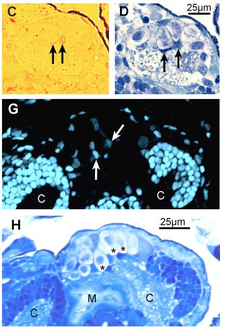
Above: (C) Two nuclei, positive for the TUNEL assay in the apical ganglion of a larva with partial velar lobes, 24 hours after induction with 5-HT. (D) Adjacent section stained with Richardson’s method. (G) Section of the apical ganglion stained with Hoechst 33342, displays no color shift (negative results) in a larva 24 hours past induction by 7-Ni. (H) Adjacent section stained with Richardson’s method.